Introduction to Head and Neck Anatomy
2. The Cranium
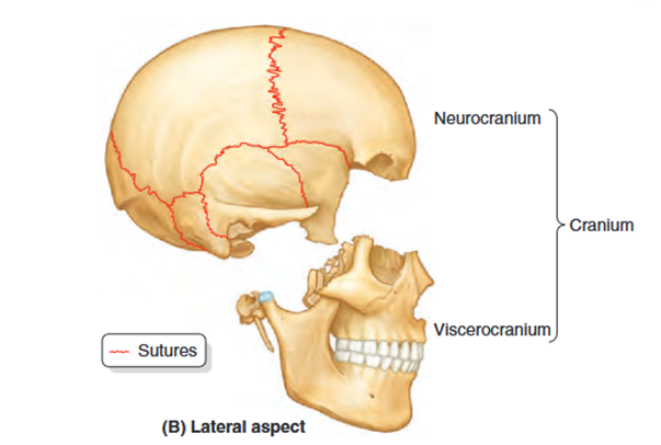
The cranium, or skull, serves as the skeleton of the head and is composed of two main parts: the neurocranium and the viscerocranium. The neurocranium encases the brain and its protective coverings, the cranial meninges, while also housing the proximal parts of cranial nerves and the brain's blood vessels. In adults, the neurocranium consists of eight bones: four singular bones aligned along the midline (frontal, ethmoidal, sphenoidal, and occipital) and two pairs of bilateral bones (temporal and parietal).
The neurocranium features a dome-like structure known as the calvaria, or skullcap, as well as a base referred to as the cranial base (basicranium). The calvaria is primarily made up of flat bones (frontal, parietal, and occipital) formed through intramembranous ossification of the head mesenchyme originating from the neural crest. In contrast, the bones that comprise the cranial base are mainly irregular bones with significant flat sections (sphenoidal and temporal), formed through endochondral ossification of cartilage (chondrocranium) or a combination of ossification types. The ethmoid bone, though irregular, contributes modestly to the midline structure.
Development of Cranium
The bones of the calvaria and some parts of the cranial base develop by intramembranous ossification. Most parts of the cranial base develop by endochondral ossification. At birth, the bones of the calvaria are smooth and unilaminar; no diploë is present. The frontal and parietal eminences are especially prominent. The cranium of a neonate is disproportionately large compared to other parts of the skeleton; however, the facial aspect is small compared to the calvaria, which forms approximately one eighth of the cranium. In the adult, the facial skeleton forms one third of the cranium. The large size of the calvaria in infants results from precocious growth and development of the brain and eyes. The rudimentary development of the face makes the orbits appear relatively large
Several bones of the cranium (frontal, temporal, sphenoid, and ethmoid bones) are pneumatized bones, which contain air spaces (air cells or large sinuses), presumably to decrease their weight. The total volume of the air spaces in these bones increases with age.
In the anatomical position, the cranium is oriented so that the inferior margin of the orbit and the superior margin of the external acoustic opening of the external acoustic meatus of both sides lie in the same horizontal plane. This standard craniometric reference is the orbitomeatal plane (Frankfort horizontal plane).