Introduction to Head and Neck Anatomy
4. Surfaces of the Skull
Facial Surface: Features of the anterior or facial (frontal) aspect of the cranium are the frontal and zygomatic bones, orbits, nasal region, maxillae, and mandible.
The frontal bone, specifically its squamous (flat) part, forms the skeleton of the forehead, articulating inferiorly with the nasal and zygomatic bones. In some adults a frontal suture persists; this remnant is called a metopic suture. It is in the middle of the glabella, the smooth, slightly depressed area between the superciliary arches.
The frontal or metopic suture divides the frontal bones of the fetal cranium into two left and right.
The intersection of the frontal and nasal bones is the nasion, which in most people is related to a distinctly depressed area (bridge of nose). The nasion is one of many craniometric points that are used radiographically in medicine (or on dry crania in physical anthropology) to make cranial measurements, compare and describe the topography of the cranium, and document abnormal variations. The frontal bone also articulates with the lacrimal, ethmoid, and sphenoid; a horizontal portion of bone (orbital part) forms both the roof of the orbit and part of the floor of the anterior part of the cranial cavity.
The supra-orbital margin of the frontal bone, the angular boundary between the squamous and orbital parts, has a supra-orbital foramen (notch) in some crania for passage of the supra-orbital nerve and vessels. Just superior to the supra-orbital margin is a ridge, the superciliary arch, that extends laterally on each side from the glabella. The prominence of this ridge, deep to the eyebrows, is generally greater in males.
The zygomatic bones also known as “cheek bones, malar bones”, forming the prominences of the cheeks, lie on the inferolateral sides of the orbits and rest on the maxillae. The anterolateral rims, walls, floor, and much of the infra-orbital margins of the orbits are formed by these quadrilateral bones. A small zygomaticofacial foramen pierces the lateral aspect of each bone. The zygomatic bones articulate with the frontal, sphenoid, and temporal bones and the maxillae.
Inferior to the nasal bones is the pear-shaped piriform aperture, the anterior nasal opening in the cranium. The bony nasal septum can be observed through this aperture, dividing the nasal cavity into right and left parts. On the lateral wall of each nasal cavity are curved bony plates, the nasal conchae.
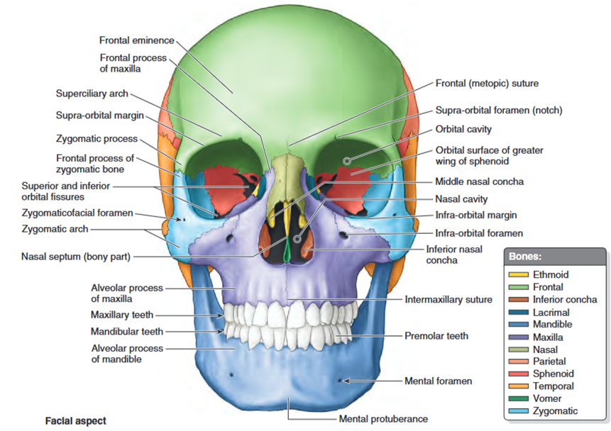
The maxillae form the upper jaw; their alveolar processes include the tooth sockets (alveoli) and constitute the supporting bone for the maxillary teeth. The two maxillae are united at the intermaxillary suture in the median plane. The maxillae surround most of the piriform aperture and form the infra-orbital margins medially. They have a broad connection with the zygomatic bones laterally and an infra-orbital foramen inferior to each orbit for passage of the infra-orbital nerve and vessels.
The mandible is a U-shaped bone with an alveolar process that supports the mandibular teeth. It consists of a horizontal part, the body, and a vertical part, the ramus. Inferior to the second premolar teeth are the mental foramina for the mental nerves and vessels
The mental protuberance, forming the prominence of the chin, is a triangular bony elevation inferior to the mandibular symphysis, the osseous union where the halves of the infantile mandible fuse.
- Key Bones: Maxilla, zygomatic, nasal, vomer, palatine, and mandible.
- Features: Includes the orbits (eye sockets), nasal aperture, and oral cavity.
- Function: Supports facial expressions, mastication, and housing of sensory organs (eyes, nose, mouth).
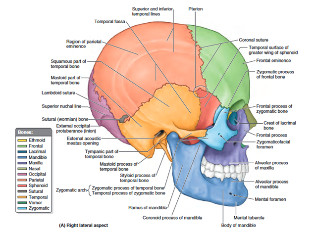
Lateral Surface
The lateral aspect of the cranium is formed by both neurocranium and viscerocranium.
The main features of the neurocranial part are the temporal fossa, the external acoustic meatus opening, and the mastoid process of the temporal bone. The main features of the viscerocranial part are the infratemporal fossa, zygomatic arch, and lateral aspects of the maxilla and mandible.
The temporal fossa is bounded superiorly and posteriorly by the superior and inferior temporal lines, anteriorly by the frontal and zygomatic bones, and inferiorly by the zygomatic arch. The superior border
- Key Bones: Temporal, parietal, frontal, and sphenoid.
- Landmarks:
Pterion: The junction of frontal, parietal, temporal, and sphenoid bones.
External acoustic meatus: Opening for the ear canal.
Zygomatic arch: Formed by the zygomatic and temporal bones.
- Clinical Note: The pterion is a vulnerable area; fractures can damage the middle meningeal artery, leading to epidural hematoma.
Occipital Surface: The posterior or occipital aspect of the cranium is composed of the occiput (back of head, the convex posterior protuberance of the squamous part of the occipital bone), parts of the parietal bones, and mastoid parts of the temporal bones.
The external occipital protuberance is usually easily palpable in the median plane; however, occasionally (especially in females) it may be inconspicuous. A craniometric point defined by the tip of the external protuberance is the inion. The external occipital crest descends from the protuberance toward the foramen magnum, the large opening in the basal part of the occipital bone.
- Key Bone: Occipital bone.
- Features:
External occipital protuberance: Palpable bony prominence.
Foramen magnum: Large opening for the spinal cord.
Occipital condyles: Articulates with the atlas (C1 vertebra).
- Function: Provides attachment for neck muscles and allows communication between the cranial and spinal cavities.
Superior External Surface
- Bones Involved: Frontal, parietal, and occipital.
- Landmarks:
Sagittal suture: Between parietal bones.
Coronal suture: Between frontal and parietal bones.
Lambdoid suture: Between parietal and occipital bones.
- Clinical anatomy: Sutures in adults are fused; in infants, they allow for skull expansion during growth.
2.5 Internal Surface of the Cranium
- Features:
Divided into three cranial fossae: anterior, middle, and posterior.
Anterior cranial fossa: Contains the frontal lobes; notable structures include the crista galli and cribriform plate.
Middle cranial fossa: Houses the temporal lobes; notable structures include the sella turcica and foramen ovale.
Posterior cranial fossa: Contains the cerebellum and brainstem; notable structures include the jugular foramen and hypoglossal canal.
Walls of the Cranial Cavity
- Structure:
Outer table: Dense, compact bone.
Diploë: Spongy bone with venous channels.
Inner table: Thin, brittle compact bone.
- Clinical anatomy: The thin inner table makes the cranial cavity susceptible to fractures and intracranial complications.