
Introduction to Head and Neck Anatomy
3. Areas of the Head
The head comprises several key areas, including the scalp, infratemporal fossa, pterygopalatine fossa, and cranial fossae. The scalp consists of skin (typically hair-bearing) and subcutaneous tissue that covers the neurocranium, extending from the superior nuchal lines on the occipital bone to the supra-orbital margins of the frontal bone. Laterally, it reaches over the temporal fascia to the zygomatic arches.
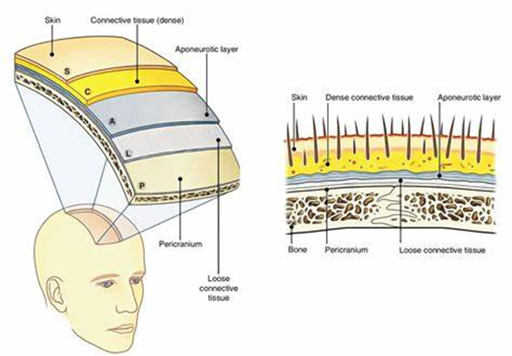
The scalp is organized into five layers, with the first three layers closely connected and functioning as a unit (e.g., when wrinkling the forehead or moving the scalp). The word "scalp" serves as a mnemonic for these layers:
- Skin: This thin layer, except in the occipital region, contains numerous sweat and sebaceous glands as well as hair follicles. It has a rich arterial supply and effective venous and lymphatic drainage.
- Connective Tissue: This thick, dense layer is richly vascularized and well-supplied with cutaneous nerves, forming the subcutaneous layer.
- Aponeurosis (epicranial aponeurosis): A broad, strong tendinous sheet covering the calvaria, it serves as the attachment point for muscle bellies from the forehead and occiput (occipitofrontalis muscle) and from the temporal bones (temporoparietalis and superior auricular muscles). Together, these make up the musculoaponeurotic epicranius. The frontal belly of the occipitofrontalis pulls the scalp forward, wrinkles the forehead, and elevates the eyebrows, while the occipital belly pulls the scalp backward, smoothing the forehead skin. The superior auricular muscle elevates the auricle of the external ear. All components of the epicranius are innervated by the facial nerve.
- Loose Areolar Tissue: This sponge-like layer contains potential spaces that can fill with fluid due to injury or infection. It allows for the free movement of the scalp proper (the first three layers) over the underlying calvaria.
- Pericranium: A dense connective tissue layer forming the external periosteum of the neurocranium. Although it is firmly attached, it can be easily stripped from the skull in living individuals, except where it is continuous with fibrous tissue in the cranial sutures.
The three outermost layers move as one unit, with the aponeurosis being a tendon-like structure spanning between the frontalis and occipitalis muscles.
The scalp receives a rich arterial supply from the external carotid artery, and sensory innervation from the trigeminal nerve, as well as cervical nerves.
The infratemporal fossa is a complex area located at the base of the skull, deep to the masseter muscles. The infratemporal fossa provides a conduit for neurovascular structures entering and leaving the cranial cavity.